The Williamsburg Bridge is a suspension bridge that spans the East River, connecting the boroughs of Brooklyn and Manhattan in New York City. An assessment of the strengths of its cables is reported in the article “Estimating Strength of the Williamsburg Bridge Cables” (R. Perry, The American Statistician, 2002:211–217). Each suspension cable consists of 7696 wires. From one of the cables, wires were sampled from 128 points. These points came from four locations along the length of the cable (I, II, III, IV). At each location there were eight equally spaced points around the circumference of the cable (A, B, C, D, E, F, G, H). At each of the eight points, wires were sampled from four depths: (1) the external surface of the cable, (2) two inches deep, (3) four inches deep, and (4) seven inches deep (the cable is 9.625 inches in radius). Under assumptions made in the article, it is appropriate to consider this as a two-factor experiment with circumferential position and depth as the factors, and with location providing four replicates for each combination of these factors. The minimum breaking strength (in lbf) is presented in the following table for each of the 128 points.
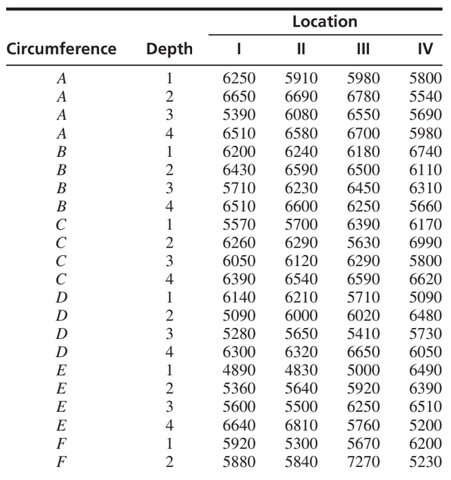
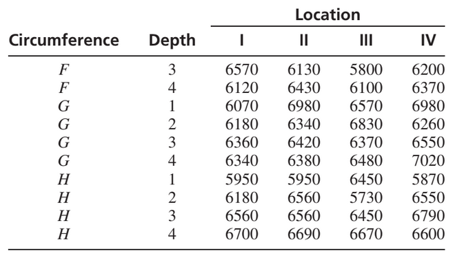
Construct an ANOVA table. You may give ranges for the P-values. Can you conclude that there are interactions between circumferential position and depth? Explain. Can you conclude that the strength varies with circumferential position? Explain. Can you conclude that the strength varies with depth? Explain.
Chapter 3: Altered Cellular and Tissue Biology Cellular Alterations Injury to cells and their surrounding environment (called the extracellular matrix) leads to tissue and organ injury Cells can adapt to physiologic demands or stress to maintain a steady state called homeostasis Adaptation is a reversible, structural, or functional response both to normal or physiologic conditions Example: uterus adapts to pregnancy—a normal physiologic process—by enlarging Example: in adverse condition such as hypertension, myocardial cells are stimulated to enlarge by increased work of pumping and are usually only temporarily successful Cellular Adaptation Physiologic vs. pathogenic o Atrophy – decrease or shrinkage in cell size and consequently in size of affected organ o Hypertrophy increase in size of cells and consequently in the size of the affected organ o Hyperplasia increase in number of cells form increased rate of cell division o Dysplasia abnormal changes in size, shape, and organization of mature cells o Metaplasia reversible replacement of one mature cell type by another, sometimes less differentiated cell type Cellular Injury Reversible Irreversible Cellular Injury Mechanisms Hypoxic injury—single most common cause of cellular injury o Ischemia—reduced blood supply o Anoxia total lack of oxygen o Cellular responses Decrease in ATP, causing failure of sodiumpotassium pump and sodiumcalcium exchange (allows calcium to enter into the cell, killing the cell) Cellular swelling Vacuolation is the formation of vacuoles within or adjacent to cells, and, in dermatopathology, often refers to the basal cellbasement membrane zone area. o Reperfusion injury restoration of oxygen that results from generation of oxygen free radicals that can cause further cell membrane damage and mitochondrial calcium overload Chemical injury o Carbon tetrachloride now banned, used in industries such as refrigeration and pesticides o Lead—paint, soil, pottery, mining o Carbon monoxide—hypoxic asphyxiation, odorless, colorless, deadly o Ethanol—alcohol intoxication/poisoning o Mercury—fish consumption and dental amalgams o Social or street drugs—heroin, meth, cocaine Unintentional and Intentional Injuries Blunt force injuries o Application of mechanical energy to the body resulting in the tearing, shearing, or crushing of tissues o Contusion vs. hematoma Contusion bruise caused by bleeding into skin or underlying tissues Hematoma is a collection of blood in soft tissue o Abrasion – wound or scrape cause by superficial damage to the skin, no deeper than the epidermis o Laceration tear or rip in tissue; ragged & irregular with abraded edges caused by blunt trauma Extreme example is an avulsion where a wide area is pulled away; organ injuries can also become lacerated from blunt force o Fractures breakage of bone from blunt force trauma Sharp injuries o Incised wounds Longer than it is deep Straight or jagged with sharp, distinct edges without abrasions Usually produces significant external bleeding and little internal bleeding May see superficial incisions in same area called “hesitation marks” o Stab wounds Penetrating sharp force injury that is deeper than it is long External bleeding small due to almost immediate tissue pressure over site o Puncture wounds Made by objects with sharp points but without sharp edges Prone to infection Can be deep (stepping on nail) o Chopping wounds Made by heavy, edged instruments such as hatchets, propeller blades Produces a combination of sharp and blunt force characteristics Unintentional and Intentional Injuries—Gunshot Wounds Contact rang entrance wounds occur when gun is held so that the muzzle rests or presses on the skin surface o Searing of edges of wounds from flame/smoke in addition to hole o Wounds is gaping and/or jagged which is called blow back that mirror the imprint of the weapon Intermediated range entrance wound: surrounded by gunpowder stippling/tattooing that results from fragments of burning or unburned pieces of gunpowder exiting gun barrel and forcibly striking skin Exit Wounds Shored exit wounds Unintentional and Intentional Injuries Asphyxia injuries—caused by a failure of cells to receive or use oxygen o Suffocation choking asphyxiation o Strangulation – hanging, ligature, and manual strangulation o Chemical asphyxiants – carbon monoxide most common, cyanide, and hydrogen sulfide (sewer gas) o drowning Infectious Injury Pathogenicity of a microorganism Diseaseproducing potential o Invasion and destruction o Toxin/endotoxin production o Production of hypersensitivity reactions Immunologic and Inflammatory Injury Phagocytic cells Immune and inflammatory substances o Histamine, antibodies, lymphokines, complement, and proteases Membrane alterations WILL DISCUSS DETAILS IN IMMUNE SYSTEM Cellular Death Necrosis: sum of cellular changes after local cell death and the process of cellular autodigestion o Coagulative necrosis Kidneys, heart, and adrenal glands Protein denaturation Usually caused by ischemia or infarction caused by chemical injury o Liquefactive necrosis Ischemic injury to neurons and glial cells of the brain and is digested by enzymes Hydrolytic enzymes break down protein, carbs, and fat Bacterial infection Staphylococci, streptococci, and Escherichia coli o Caseous necrosis Tuberculous pulmonary infection Combination of coagulative and liquefactive necrosis Cottage cheese appearance and is soft and granular o Fat necrosis Breast, pancreas, and other abdominal organs Action of lipases break down fat in these organs o Gangrenous necrosis Death of tissue from severe hypoxic injury commonly from arteriosclerosis or blockage of major arteries, esp. those in legs Dry gangrene dries and shrinks skin and color turns to dark brown or black Wet gangrene develops when WBCs invade the site, usually internal organs causing site to become cold, swollen, and back with foul odor Gas gangrene refers to special type of gangrene caused by infection of injured tissue by the bacteria clostridium Death is caused by shock Apoptosis (programmed cell death) vs. necrosis Aging Cellular aging o Atrophy, decreased function, and loss of cells Tissue and systemic aging o Progressive stiffness and rigidity o Sarcopenia—loss of muscle tissue R/T aging Frailty o Mobility, balance, muscle strength, motor activity, cognition, nutrition, endurance, falls, fractures, and bone density Somatic Death Algor mortis—post mortem reduction of body temperature; takes about 24 hours until the body temperature equals that of the environment Livor mortis—gravity causes blood to settle in the most dependent or lowest tissues which develop a purplish discoloration Rigor mortis—occurs over 6 14 hours; muscle stiffening with smaller muscles being affected first Putrefaction usually occurs between 2448 hours after death as rigor mortis gradually diminishes Body becomes flaccid in 3662 hours